|
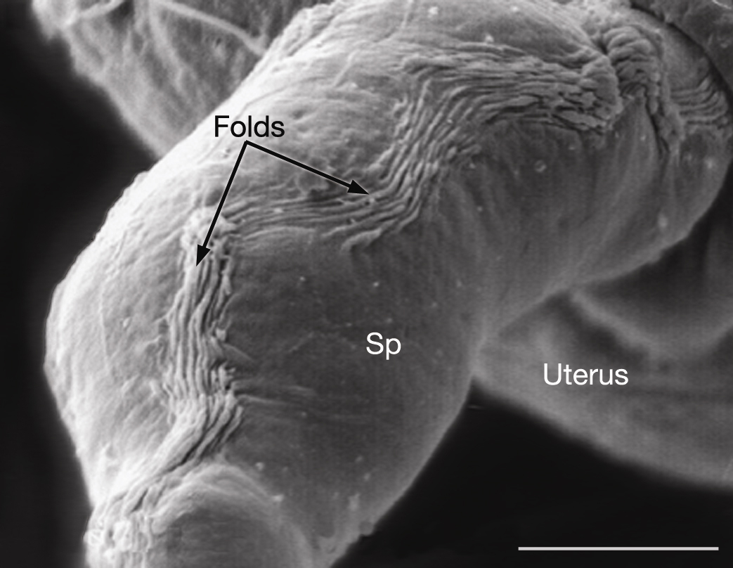
EMSEMDissectionFIG 2: Spermatheca viewed by SEM after dissection.
SEM, dorsolateral view of an adult (dissected) gonad, proximal arm showing outer surface of the epithelium contracting into parallel folds when not inflated to enclose a primary oocyte. The spermatheca (Sp) is empty of oocytes. Scale bar,
6.1 μm. (Image source: L. Hoffman and D. Greenstein, photo SP4.)
Click on picture for full resolution image.
|